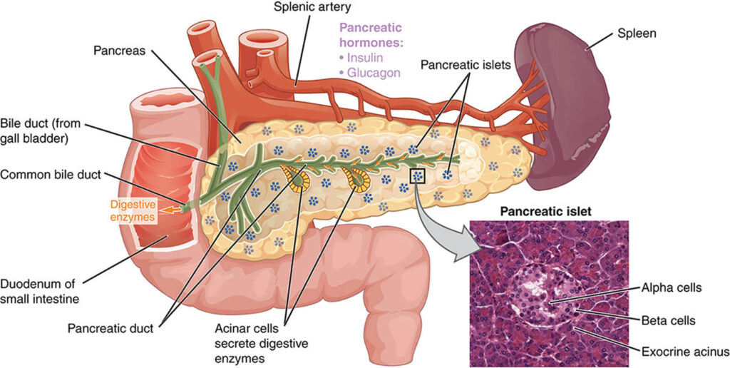
The pancreas, named for the Greek words “pan” (all) and “kreas” (flesh), is a 12-15–cm long comma-shaped, soft, lobulated, retroperitoneal organ. It lies transversely, although a bit obliquely, on the posterior abdominal wall behind the stomach, across the lumbar (L1-2) spine.
Gross Anatomy
The pancreas is arbitrarily divided into head, uncinate process, neck, body and tail. The pancreatic head constitutes about 50% and the body and tail the remaining 50% of the pancreatic parenchymal mass. The pancreas weighs about 80gms.

The pancreas is a prismoid in shape and appears triangular in cut section with superior, inferior, and anterior borders as well as anterosuperior, anteroinferior, and posterior surfaces. On the cut surface of the pancreas at its neck, the main pancreatic duct lies closer to the superior border and the posterior surface.
The head of the pancreas lies in the duodenal C loop in front of the inferior vena cava (IVC) and the left renal vein. The uncinate process is an extension of the lower (inferior) half of the head toward the left; it is of varying size and is wedged between the superior mesenteric vessels (vein on right, and artery on left) in front and the aorta behind it.
The lower (terminal) part of the common bile duct runs behind (or sometimes through) the upper half of the head of pancreas before it joins the main pancreatic duct of Wirsung to form a common channel (ampulla), which opens at the papilla on the medial wall of the second part of the duodenum.
The neck of the pancreas lies in front of the superior mesenteric vein, splenic vein and portal vein junction. The body and tail of the pancreas run obliquely upward to the left in front of the aorta and left kidney. The pancreatic neck is the arbitrary junction between the head and body of the pancreas. Portal vein lies behind the neck of the pancreas; no tributaries drain from the posterior surface of the pancreas into the anterior surface of the portal vein; therefore, a tunnel can be easily created behind the neck of the pancreas before its division. The narrow tip of the pancreas tail reaches the splenic hilum in the splenorenal (lienorenal) ligament.
The duodenum (25 cm long) is horseshoe-shaped, with its inferior limb longer than the superior, and has 4 parts: (1) superior (5 cm) at the level of L1; (2) descending, or C loop (7.5 cm), at L1-L3; (3) horizontal, or transverse (10 cm), at L3; and (4) ascending (2.5 cm), leading to the duodenojejunal flexure (junction).
The transverse mesocolon (with the middle colic vessels in it) is attached to the anterior surface of the lower (inferior) part of body and pancreas tail; thus, most of the gland is located in the supracolic compartment. The body and tail of the pancreas lie in the lesser sac (omental bursa) behind the stomach.
Blood supply
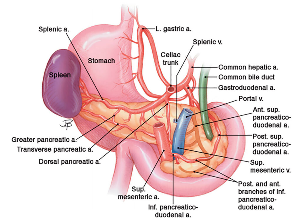
Pancreas derives a rich blood supply from both celiac axis and superior mesenteric artery, with collaterals between the two systems; that is why when angiography is done for bleeding as a complication of acute pancreatitis, chronic pancreatitis or pancreatoduodenectomy both celiac axis and superior mesenteric artery should be evaluated.
The celiac trunk (axis) comes off from the anterior surface of the aorta at the level of T12–L1. It has a short length of about 1 cm and trifurcates into the common hepatic artery (CHA), splenic artery, and left gastric artery (LGA). The CHA runs toward the right on the superior border of the proximal body of the pancreas, and the splenic artery runs toward the left on the superior border of the distal body and tail of the pancreas.

The superior mesenteric artery (SMA) comes off from the anterior surface of the aorta just below the origin of the celiac trunk at the level of L1 behind the neck of the pancreas. Then, it descends down in front of the uncinate process and the third (horizontal) part of the duodenum to enter the small bowel mesentery.
The gastroduodenal artery (GDA), a branch of the CHA, runs down behind the first part of the duodenum in front of the neck of the pancreas and divides into the right gastro-omental (gastroepiploic) artery (RGEA) and superior pancreaticoduodenal artery (SPDA), which further bifurcates into anterior and posterior branches. The inferior pancreaticoduodenal artery (IPDA) arises from the SMA and also bifurcates into anterior and posterior branches.
The anterior and posterior branches of the SPDA and IPDA join each other and form anterior and posterior pancreaticoduodenal arcades in the anterior and posterior pancreaticoduodenal grooves supplying small branches to the pancreatic head and uncinate process of the pancreas as well as the first, second, and third parts of the duodenum (vasa recta duodeni). Multiple pancreatic branches (including a dorsal pancreatic artery, great pancreatic artery or arteria magna pancreatica) of the splenic artery supply the pancreatic body and tail. Multiple, small pancreatic branches of a dorsal pancreatic artery from the splenic artery and an inferior pancreatic artery from the superior mesenteric artery supply the body and tail of pancreas.
The arterial supply of the pancreas forms an important collateral circulation between the celiac axis and superior mesenteric artery.
Veins accompany the SPDA and IPDA. Superior pancreaticoduodenal veins (SPDVs) drain into the portal vein and inferior pancreaticoduodenal veins (IPDVs) drain into the superior mesenteric vein (SMV). A few small, fragile uncinate veins drain directly into the SMV. Some veins from the head of the pancreas drain into the gastrocolic trunk. Numerous small, fragile veins drain directly from the pancreatic body and tail into the splenic vein.
The SMV lies to the right of the SMA in front of the uncinate process and the third part of the duodenum. The splenic vein arises in the splenic hilum behind the tail of the pancreas and runs from left to right on the posterior surface of the pancreatic body. Union of the horizontal splenic vein and the vertical SMV forms the portal vein behind the neck of the pancreas.
The inferior mesenteric vein (IMV) joins the splenic vein (or the junction of the splenic vein and SMV, or even SMV). The portal vein receives the SPDVs, right gastro-omental (gastroepiploic) vein, left gastric vein (LGV), and right gastric vein (RGV); then, it runs up (superiorly) behind the first part of the duodenum in the hepatoduodenal ligament behind (posterior to) the common bile duct on the right and proper hepatic artery on the left. The portal venous system (splenic vein, SMV, and portal vein) has no valves.

Lymphatic drainage
The head of the pancreas drains into pancreaticoduodenal lymph nodes and lymph nodes in the hepatoduodenal ligament, as well as pre-pyloric and post-pyloric lymph nodes. The pancreatic body and tail drain into mesocolic lymph nodes (around the middle colic artery) and lymph nodes along the hepatic and splenic arteries. Final drainage occurs into celiac, superior mesenteric, and para-aortic and aortocaval lymph nodes.
Nerve supply
The pancreas receives parasympathetic nerve fibers from the posterior vagal trunk via its celiac branch. Sympathetic supply comes from T6-T10 via the thoracic splanchnic nerves and the celiac plexus.


